
Prolapse of the uterus may occur in any species; however, it
is most common in dairy and beef cows
and ewes and less frequent in sows. Prolapse of the uterus invariably occurs
immediately after or within several hours of parturition, when the cervix is open
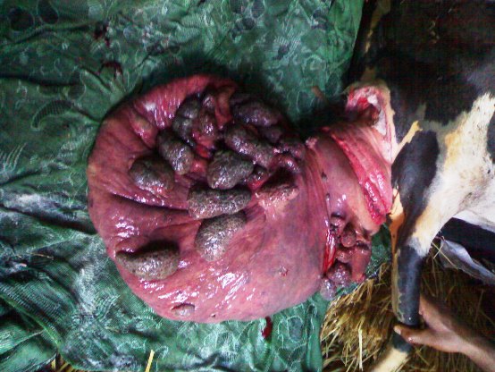
and the uterus lacks tone. Prolapse of the post gravid uterine horn usually is
complete in cows, and the mass of uterus usually hangs below the hocks. The
invagination of the contralateral horn can be located by careful examination of
the surface of the prolapsed organ.
The cause is unclear and occurrence is sporadic. Following are the contributory causes.
- Recumbence with the hindquarters lower than the forequarters
- Invagination of the tip of the uterus
- Excessive traction to relieve dystocia or retained fetal membranes
- Uterine atony
- Hypocalcemia
- Lack of exercise
In cows, treatment involves removing the placenta, thoroughly cleaning the endometrial surface, and repairing any lacerations. Rubbing the surface of the uterus with glycerol helps reduce edema and provides lubrication. The uterus is then returned to its normal position. An epidural anesthetic should be administered first. If the cow is standing, the cleansed uterus should be elevated to the level of the vulva on a tray or hammock supported by assistants, and then replaced by applying steady pressure beginning at the cervical portion and gradually working toward the apex.
Once the uterus is replaced, a hand should be inserted to the tip of both uterine horns to be sure that there is no remaining invagination that could incite abdominal straining and another prolapse. Installation of warm, sterile saline solution is useful for ensuring complete replacement of the tip of the uterine horn without trauma. If recumbent, the cow should be positioned with the hindquarters elevated by placing her in sternal recumbency with the hind legs extended backward.
Each handling in the field, we strive in accordance with that recommended in veterinary medical, because we realize that we live in a small area and veterinary medical equipment are extremely limited. As in this case, we use simple tools and used a modified retaining vulva of a toothbrush body. During one week, cow is placed with high posterior position and after one week, the appliance should be removed, after confirmed by rectal examination to know the recovery process in the uterus.
The uterus is out! Way to put it back in!
After giving birth the uterus of this cow came out because of contractions. This is the way to put it back in and with medicine the cow recovered satisfactorily.
The prognosis depends on the amount of injury and contamination of the uterus. The prognosis is favorable when a clean, minimally traumatized uterus is promptly replaced. There is no tendency for the condition to recur at subsequent parturitions. Complications tend to develop when laceration, necrosis, and infection occur, or when treatment is delayed. Shock, hemorrhage, and thromboembolism are potential sequelae of a prolonged prolapse.
Treatment of cervico-vaginal
prolapse in cow
In some instances, the bladder and intestines may prolapse into the everted uterus. These require careful replacement before the uterus is replaced. The bladder may be drained with a catheter or needle passed through the uterine wall. Elevation of the hindquarters and pressure on the uterus aids in replacement of bladder and intestines. It may be necessary to incise the uterus carefully (in a longitudinal direction) to replace these organs. In cows, amputation of a severely traumatized or necrotic uterus may be the only means of saving the animal. Supportive treatment and antibiotic therapy are indicated.
No comments:
Post a Comment