 Background
Canine Babesiosis is a
protozoan disease caused by Babesia canis or Babesia gibsoni and is transmitted
by ticks to susceptible canine hosts. It is one-celled organisms in red blood cells. This disease is sometimes associated with
other tick-borne diseases, such as Lyme disease, canine Ehrlichiosis, and Rocky
Mountain spotted fever, among others. This can make symptoms worse and
complicate diagnosis.
Canine Babesiosis occurs
worldwide, mostly in regions where ticks are prevalent. Young pets tend to
become infected most often, and with worse symptoms.
Pathogenesis
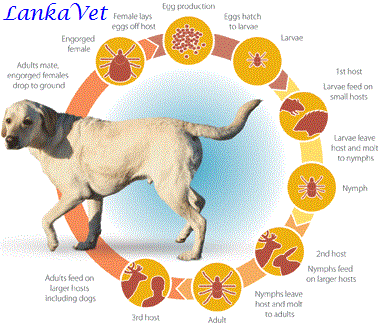 Animals are affected after an
infected tick bites and feeds on a susceptible host for a minimum of three
days. When the Babesia organism is
introduced into the host, it attaches to red blood cell membranes and is penetrated. Hemolytic anemia and hypotensive shock are
typical clinical syndromes of infection.
Hemolytic anemia results from direct erythrocyte damage by the parasite,
and both intravascular and extravascular immune-mediated destruction of red
blood cells.
Animals are affected after an
infected tick bites and feeds on a susceptible host for a minimum of three
days. When the Babesia organism is
introduced into the host, it attaches to red blood cell membranes and is penetrated. Hemolytic anemia and hypotensive shock are
typical clinical syndromes of infection.
Hemolytic anemia results from direct erythrocyte damage by the parasite,
and both intravascular and extravascular immune-mediated destruction of red
blood cells.
Clinical Signs & Symptoms
- Loss of appetite
- Lethargy
- Fever greater than 105.8℉
- Hemolytic anemia - Pale tongue, gums, and nose due to severe deficiency of red blood cells
- Hematuria - Red or orange color urine
- Enlarged lymph nodes
- Icterus or jaundice - Yellowing of the skin and the whites of the eyes caused by an accumulation of bile pigment (bilirubin) in the blood
- Splenomegaly & Lymphadenomegaly

Diagnosis
Diagnosis begins with a
complete history and a physical exam. Your veterinarian will most likely do the
following:
History
- Your veterinarian will likely inquire about recent tick exposure and bite
history.
Complete Blood Count
- These blood tests will evaluate various internal organ functions, including
the heart, liver, kidneys, pancreas, metabolism, and electrolyte balance. The
CBC is a measure of the amount and different kinds of red and white blood cells
that are present in your dog’s body.
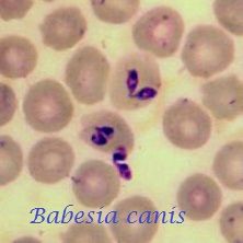
 Blood Smear
- This technique is used to examine the individual cells in your dog’s blood. Infection
with B. canis or B. gibsoni is definitively diagnosed by demonstration of the parasites
on red cells. Your veterinarian will use a drop of blood from your dog’s leg or
neck, and spread it thin on a slide. They will then examine the slide under a
microscope in order to see the Babesia parasite.
Blood Smear
- This technique is used to examine the individual cells in your dog’s blood. Infection
with B. canis or B. gibsoni is definitively diagnosed by demonstration of the parasites
on red cells. Your veterinarian will use a drop of blood from your dog’s leg or
neck, and spread it thin on a slide. They will then examine the slide under a
microscope in order to see the Babesia parasite.
Immunofluorescence
- This is a technique used to illuminate a pathogen or antibodies to a pathogen
in a tissue or culture using a fluorescent dye. In this case, your veterinarian
will make a tissue or cell smear and expose it to a specific antibody for Babesiosis.
The dye will attach to any Babesia antibody displayed in the sample and show
under a microscope as a bright green spot on the slide.
Treatment
 Following are the most
effective drugs used in the treatment of canine Babesiosis.
Following are the most
effective drugs used in the treatment of canine Babesiosis. - Diminazene Aceturate,
- Imidocarb Dipropionate
- Clindamycin - Considered in refractory or more severe and virulent infections.
Prevention
Prevention of canine Babesiosis
is mostly aimed at controlling the vector.
The environment should be
treated to decrease tick numbers, dogs should be treated to control tick
infestations, and ticks should be removed from parasitized animals as quickly
as detected.
 Recently, a vaccine which
minimizes the severity of infection was developed. The vaccine is reported to be 70 to 100%
effective in diminishing the pathologic effects which typically ensue upon
infection.
Recently, a vaccine which
minimizes the severity of infection was developed. The vaccine is reported to be 70 to 100%
effective in diminishing the pathologic effects which typically ensue upon
infection.
Blood transfusion poses a
significant risk to recipient animals; therefore it is recommended that donor
animals be tested for infection with Babesia organisms.
Conclusion
It is important for
practitioners to keep common diseases such as Babesiosis in the list of
differential diagnoses for acute hemolytic anemia, shock and icterus.
very useful
ReplyDelete