Saturday, February 2, 2013

Roy's First Birth Day


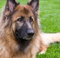
        This is my dog; Roy.  It is one year old. His birth day was 02.02.2012. Today is Roy’s first birthday. This post is written to celebrate his first birthday. Roy is a crossbred German Shepherd dog. His mother is totally a mongrel, but father had good German shepherd features.


I brought him to my home, when he was one and half months old. Having traveled in my arms for some time from the breeder’s premises, he was bit nervous when reaching to home. 


After some time of arrival to home, we bathed him as he smells bit bad. We then directly taken him to our bed room and placed him on the bed and we were watching for some time, what he was trying to do. He was bit reluctant move and looks very timid.  Roy was given some milk to drink, he was bit happy now. We have been much exiting with the arrival of Roy and did every possible things to get familiar with him. We gave him the name; Roy, and tried to convince it being called his name. It is a short name which everyone can pronounce in the same way.
 In the first few hours at our home, Roy had begun learning to use the new environment and he got used to come towards us when called his name; Roy.


We sometimes call him Roy de Shane for fun, as his father’s name is Shane. Roy spent his first night in our bed room, until the puppy is really familiar with a proper sleeping place. After two weeks time, he was introduced a new cage where he was reluctant to live in, but in a very few days, he got used to it. Roy is very much like to stay beside us or hang out in our bedroom.
Roy's body weight was measured after one month of arrival and it was 5 kg.


       After some time, we introduce him a friend who is our little female cat; Kitty. Kitty was six months older than Roy and bit holistic towards him. Initially; Kitty refused to getting friendly with Roy as she was bit frightened. With the time, she got familiarized with Roy and play a little bit.
         Frequently, we pick it up and let others in our family to do so. He did not struggle much when he is in our arms.


When Roy was three and half months, we measured his body weight again, and it was 8.25 kg. He was sitting on the weighing balance very cute way trying to balance himself on it.


When Roy is six months old, his body weight was 18 kg. Now, he is much mischievous and he often wants to play with us. 


Roy was given a very good veterinary care including vaccination against the all infectious diseases and proper deworming. His body weight was measured monthly and bathed him in every other  day as he was often trying to get in our bed. He is very much like to have a bath.


When he was six months old, he got infected with tick fever and I treated him accordingly; but, after two weeks time, he got the second attack again. Having cured within a few days time, he was well treated against tick infestation and his cage and surrounding area was disinfected, then detickings was spreaded.




Thursday, January 31, 2013

Dancing Dogs


Abscesses in your Pets


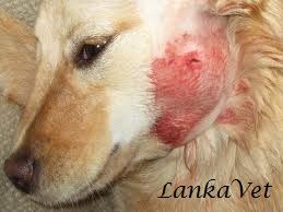
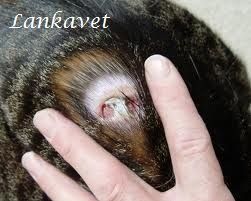
An abscess is a collection of pus encapsulated within a tissue underneath the skin. It occurs when the body tries to fight an infectious process usually caused by bacteria or parasites within the body tissues, sending white blood cells to fight further infection.
An abscess may first appear as a painful firm swelling under the skin that is hot or cold to the touch. In later stages, the localized swelling often becomes softer and less painful. This usually happens right before the abscess is getting ready to burst and drain.
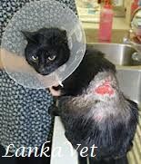
Abscesses around the neck and tail are far most common in cats who have access to the outdoors and who play territorial games with their fellow cats. Their teeth and claws are extremely contaminated with bacteria, and when they are sunk into the skin of the opposing cat, the bacteria becomes embedded under the skin. Pasteurella multocida is the most common bacteria to cause skin infections.

Causes

The most common causes of abscesses include:
  • Animal bites
  • Scratches
  • Trauma to the skin or underlying tissues
  • Foreign object penetration
  • Bad oral hygiene


How does it occur?

For an abscess to occur, the pet must have been injured somehow. They are caused by obstruction of oil (sebaceous) glands or sweat glands, inflammation of hair follicles, or minor abrasions and punctures of the skin. Germs get under the skin or into these glands, which causes an inflammatory response as your pet's natural defense mechanisms kick-in to remove the foreign body.
The middle of the abscess liquefies and contains dead cells, bacteria, and other debris. This area begins to grow, creating tension under the skin and further inflammation of the surrounding tissues. Pressure and inflammation cause the pain.
Pus is formed when the white blood cells die and accumulate in the affected area. As a protective mechanism, the immune system also works to wall off this area from the rest of the body, by forming a fibrous capsule. This fibrous capsule keeps the pus confined to one area. If the bacteria or foreign material overwhelms the immune system, the pus continues to accumulate and the abscess becomes larger.
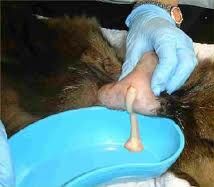
As time goes on, the wall surrounding the ever-growing pus pocket becomes thin, and at some point, it ruptures and pus drains out. Typically, the abscess ruptures to the exterior of the body, and pus drains from the wound. The pus is usually quite thick and creamy, and often has a distinct smell. However, in many cases; the abscess may not drain naturally and must be excised by a vet.

Signs
  • Swelling and inflammation
  • Heat and redness around the area
  • Pain around the area of infection
  • High temperature or fever
  • Lack of appetite
  • Lethargy and a reluctance to move
  • Rotting teeth, tooth loss, excessive saliva and bad breath (in mouth abscesses)
  • Hair loss (in skin abscesses)
  • Stiffness or lameness (abscesses around joints)
  • Licking or grooming affected area excessively
  • Foul odor
  • Purulent discharge (pus) from a wound

Treatment
  • Abscess drainage is painful and better to do it under anesthesia
  • Painkillers
  • Antibiotics to avoid secondary bacterial infections - Topical & intramuscular
  • Clindamycin - If it is serious or deep in the skin

Caring and Management
  • Clean hands thoroughly before handling abscess.
  • Clear the surrounding skin by clipping away hair with a small pair of scissors.
  • Gently clean the area with a mild soap and then rub it with clean gauze soaked with antibacterial solution, Ex. saline solution or hydrogen peroxide.
  • Gently pat the area dry with a towel.
  • Drain the abscess via surgical puncture and then drain with a sterile solution such as normal saline.
  • Abscesses are usually left open and allowed to drain to prevent the re-infection. 
  • Use a topical skin protecting agents, such as Betadine, to guard the skin against bacterial infection and keep the affected area clean.
  • Check it every 12 hours. Gently remove any scabs or crusts skin that have formed and clean the area with soap and water apply Betadine ointment.
  • If you come in contact with draining pus, just wash your hands. These bacteria may infectious to humans if it is exposed to an open wound on skin.

Sunday, January 27, 2013

Chopsticks Feeding to Kitten


ThumbnailTo avoid being bitten and pounced on during kitty training, we have been using chopsticks and toothpicks to feed the kittens their treat. As seen in this video, this technique has worked particularly well with training "Gluteus," where the kittens will sit up straight, balancing on their buttocks muscles. 

The gluteus maximus is the largest and most superficial of the three gluteal muscles. It makes up a large portion of the shape and appearance of the buttocks.

Exocrine Pancreatic Insufficiency - EPI


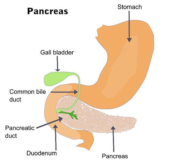
Exocrine pancreatic insufficiency (EPI) is a syndrome of maldigestion and malabsorption, caused by insufficient synthesis and secretion of digestive enzymes by the exocrine portion of the pancreas. It is much more common in dogs than cats. It is also referred to as Pancreatic Hypoplasia or Pancreatic Acinar Atrophy (PAA).
Pancreas has two primary functions;
  • Endocrine cells produce and secrete hormones, insulin, and glucagon.
  • Exocrine cells produce and secrete digestive enzymes.

 
Causes of this disease

EPI is the inability of the pancreas to secrete digestive enzymes: amylase to digest starches, lipases to digest fats, and proteases to digest protein. Pancreatic acinar atrophy is the most common cause of EPI in dogs, while chronic pancreatitis is the most common cause in cats. Less common causes of EPI in dogs and cats are pancreatic or extra-pancreatic masses that lead to obstruction of the pancreatic duct.
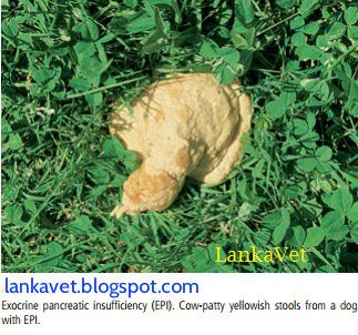
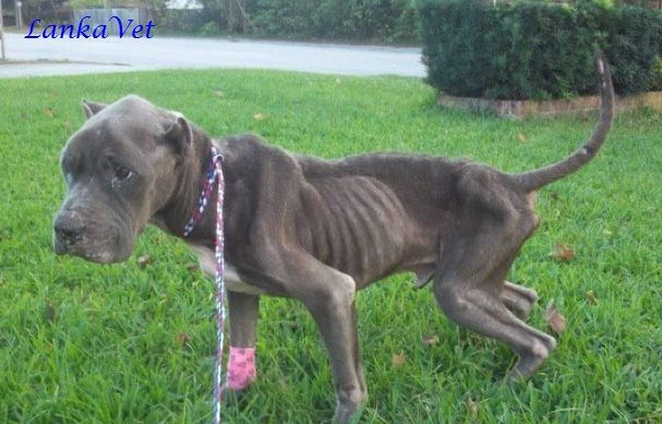
Lack of pancreatic digestive enzymes may lead to maldigestion & malabsorption, therefore; the nutrients remaining in the intestinal lumen lead to loose, voluminous stools and steatorrhea (excess fat in stool).  It also causes weight loss and may lead to vitamin deficiencies. Due to the lack of absorbed nutrients, the body starves: muscle mass wastes away, and bones may also be affected.
Clinical signs
  • Voracious appetite - Polyphagia  
  • Rapid Weight loss
  • Intermittent watery diarrhea or vomiting
  • Eating stools – Coprophagia
  • Eating non-food items - Pica
  • Dry, dull, or brittle coat
  • Feces is pale or grayish, loose, and voluminous and may be malodorous
  • Increased rumbling sounds from the abdomen, and passing increased amounts of gas
  • High fat content of the feces can lead to a greasy appearance of the hair coat, especially in the perianal and tail region of cats.
  • Temperament changes showing fear or aggression

 
Diagnosis
  • Clinical signs
  • Fecal Protease Test - Determining the levels of digestive enzymes in the stool
  • Serum Trypsin-like Immunoreactivity Test- Determining the levels of certain digestive enzymes in the blood


Treatment
  • Treatment involves replacing the dog's pancreatic enzymes: Freeze-dried and ground-up extracts of pig and cattle pancreases could be given. Accurate dosing is more difficult with the raw pancreas, but in general, a 45-pound dog would need to receive 3 to 4 ounces of the chopped pancreas. The raw pancreas can be frozen at 4ºF for up to 3 months and still retain its enzyme activity. They can also be formulated either into tablets or powder.  Ex.  Viokase-V,  Pancreatin 6X,  Bio Case V. Powder is more effective than tablets, capsules, and especially enteric-coated products. Initially, 2 tsp/20 kg body wt should be given with each meal for dogs and 1 tsp/cat with each meal for cats.
  • Concurrent antacid therapy has little effect on overall digestive ability
  • Oral vitamin E supplementation or intra muscular injections of vitamin B-12
  • A low fat, low fiber, highly digestible diet

Friday, January 25, 2013

Mastitis in Cows


Mastitis is an inflammatory reaction of mammary glands caused by various infectious or non-infectious etiological agents. It is characterized by a series   of physical, chemical and microbiological changes and some pathological changes in the mammary tissue and udder.  Consequently it causes increase in the number of somatic cells in the milk, changes in milk composition and reductions in milk yield. Adult lactating cows are most at risk for infections.

The infection occurs when the micro organisms gain access to the udder through the teat canal. The infections in the udder affect the quantity and quality of the secreted milk. The disease is known to cause economic losses of up to 40 % of the herd productivity.

However, mastitis is a global problem as it adversely affects animal health, quality of milk and economics of milk production and every country including developed ones suffer huge financial losses.

Mastitis can be described as;
  • Clinical mastitis
  • Subclinical mastitis


Clinical Mastitis

The inflammatory response to the infections of clinical mastitis is visible and the abnormal milk is readily detected.

Subclinical Mastitis
  • No changes in the milk is apparent.
  • No obvious clinical signs such as abnormal milk, udder swelling or tenderness, or systemic signs.
  • It can only be detected by the laboratory examination of milk drawn from the udder of affected cow.


Pathogens involved

  • Streptococcus agalactiae
  • Streptococcus dysgalactiae
  • Streptococcus uberis
  • Staphylococcus epidermidis
  • Staphylococcus aureus
  • Enterococci
  • Corynebacterium sp
  • Klebsiella sp.
  • Pseudomonas aeriginosa
  • Brucella meletensis
  • Mycoplasma sp
  • Pasturella sp
  • Proteus

Common causes and transmission
  • Mastitis is usually caused by poor sanitation in the sheds like unclean floorings, bedding, animals and the milking surroundings. Unhygienic conditions favor multiplication of bacteria and infect the animals.
  • Poor milking practice, not washing the animal’s udders before milking, not practicing the dipping of teats in antiseptic solution and incomplete milking of animals are also responsible for mastitis.
  • Infection also spreads through milking machines if they are not cleaned well before use.
  • Infection also spreads through milking persons if their hands are contaminated and not cleaned well with a disinfectant before milking.
  • Improper milking methods like milking using thumb and pressing or pulling the teat causes a mechanical injury to the teat.
  • Injuries to the udder or teats also cause mastitis.


Symptoms
  • Redness and pain of udder
  • Swelling of udder and teats
  • Milk gets curdled becomes watery with blood streaks in some cases
  • Fever, listlessness and anorexia
  • Reduced milk production
  • In chronic and sub clinical cases the teat canal gets thickened, when the teat is palpated it appears like a thread inserted in the teat canal.
  • In subclinical cases, the milk is not affected and appears normal but the somatic cell count increases and the milk yield are decreased. If the subclinical cases are more in the farm it becomes a problem to control the disease.

Diagnosis

Based on clinical signs;
  • Strip cup test to detect the curdled milk pieces
  • California Mastitis Test
  • Milk culture to identify the causative organism


Treatment

Intramammary infusions of antibiotic: Prior to intramammary infusion, the teat is cleaned well and the tip of the teat is swabbed with an alcohol swab and allowed to dry for a number of seconds. The antibiotic comes in a plastic tube with a plastic infusion cannula on the end.

  • Penicillin at a dose rate of 50 - 200,000 units
  • Tetracycline at a dose rate of 100 - 400 mg
  • Streptomycin at a dose rate of 0.25 - 1 g
  • Neomycin at a dose rate of 0.5 - 1 g
  • Polymixin at a dose rate of 50 mg
  • Erythromycin at a dose rate of 300 - 600 mg
How to apply antibiotic directly into the teat:
Step 1: Milk the udder until it's empty                
Step 2: Clean the end of the teat
Step 3: Put the tip of the tube into the
              teat and squeeze the antibiotic
              up into the udder  
Step 4: Massage the teat and the udder

Oxytocin treatment: 
A key contributing factor to effective milk let down. Clearly removal of the primary growth medium of the bacteria, the milk, more often should enhance rate of recovery from infection.

Wednesday, January 23, 2013

Parvo Virus Infection in Dogs


The canine parvovirus (CPV) infection is an acute, highly contagious viral disease that affects dogs. CPV has a tendency to attack rapidly reproducing cells, such as those lining the gastrointestinal tract. CPV affects dogs of all ages, but the majority of cases are seen in puppies that are between six weeks and six months old. Doberman Pinschers and Rottweilers appear to acquire the infection more readily and experience more severe symptoms. The reason for lower resistance in these breeds is unknown.

This virus manifests in two different forms.
  •        Intestinal form - more common
  •        Cardiac form     - less common


Transmission

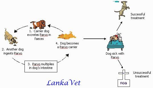
CPV is transmitted either by direct contact with an infected dog, or indirectly, by the fecal-oral route.
The virus sheds in large amounts in the stools of acutely infected dogs for up to several weeks following infection. This virus can be carried on the dog’s hair and feet, as well as on contaminated crates, shoes, and other objects. When the dog licks the fecal material off hair, feet, or anything that came in contact with infected feces, he acquires the disease.

Clinical signs

Intestinal form:
  • Anorexia- Loss of appetite
  • Lethargy
  • Fever - 106°F
  • Vomiting
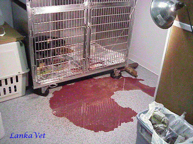
  • Severe bloody diarrhea
  • Heavy weight loss
  • Dehydration
  • Mucous membranes become red
  • Abdominal pain or discomfort on palpation
  • Increased heart beat


Cardiac form:
  • Attacks the heart muscles of very young puppies
  • Sudden death
  • Crying, difficulty in breathing, gasping for breath
  • Extreme depression
  • Weakness
  • Unwillingness to nurse
  • Irregular heartbeat


  Diagnosis
  It is based on;
  •     Physical examination of clinical signs
  •     Biochemical tests
  •     Urine and fecal analysis
  •     Abdominal radiographs
  •     Abdominal ultrasounds

Treatment

No treatment found as the disease is a viral infection. Supportive treatment is focused on curing the symptoms and preventing secondary bacterial infections.
  • Intravenous fluid and nutrition therapy is crucial in maintaining normal body fluid after severe diarrhea and dehydration
  • Antiemetics – to stop vomiting
  • H2 Blockers to reduce nausea
  • Antibiotics – to counteract secondary bacterial infections
  • Anthelmintics – to kill intestinal worms


Management after recovering

Even after your dog has recovered from a CPV infection, it will still have a weakened immune system, and will be susceptible to other illnesses. Therefore; you should boost your dog's immune system.  A diet that is easily digested will be best for your dog while it is recovering.
Your dog will also continue to be a contagion risk to other dogs for at least two months after the initial recovery. You will need to isolate your dog from other dogs for a period of time. Wash all of the equipments that your dog uses (e.g., dishes, kennel, toys) with non-toxic cleaners. Recovery comes with long-term immunity against the parvovirus, but it is no guarantee that your pet will not be infected with the virus again.

Prevention

Thoroughly clean and disinfect the cages of infected animals. Parvo is an extremely hardy virus that resists most household cleaners and survives on the premises for months. The most effective disinfectant is household bleach in a 1:32 dilution. The bleach must be left on the contaminated surface for 20 minutes before being rinsed.

Vaccinations:

·      Start by 6 weeks of age,
During the first weeks of life, puppies are protected by high levels of maternal antibodies. As these levels decline gradually, from 2-4 weeks puppies are more susceptible to infection.
·  Booster vaccination is given at 12 weeks of age and thereafter, annual re-vaccination should be given.