Sunday, February 24, 2013

Toxoplasmosis in Cats


Toxoplasmosis is a disease caused by a single-celled intracellular parasite called Toxoplasma gondii. It is a protozoan parasite which can infects multiple of warm blooded animals including humans, livestock, birds and especially cats. However, cats are the definitive hosts in which the parasite is only able to sexually reproduce in cats.


Causes

The life cycle of Toxoplasma gondii has two hosts;
  • Definitive host - cats
  • Intermediate host – other domestic animals & humans


Parasite can only produce eggs when infecting a cat. When a cat ingests an infected prey or infected raw meat the parasite is released into the cat's digestive tract. The organisms then multiply in the wall of the small intestine and produce eggs. These eggs are then excreted in large numbers in the cat's stool.

Symptoms
  • Loss of appetite
  • Lethargy
  • Weight loss
  • Abdominal Pain
  • Vomiting
  • Diarrhea
  • Depression
  • Fever
  • Shortness of breath
  • Uncoordinated gait
  • Seizures
  • Tremors
  • Muscular weakness
  • Partial or complete paralysis
  • Jaundice
  • Inflammation of tonsils - tonsillitis
  • Inflammation of retina - retinitis
  • Inflammation of middle part of the eye including iris - uveitis
  • Inflammation of the cornea – keratitis

Symptoms are most severe in kittens infected while in the womb. These kittens may be stillborn or die before weaning. Those which survive may show lack of appetite, fever, difficulty in breathing, and jaundice


Diagnosis

Based on;  

* History
* Clinical signs
* Laboratory tests: Determine the   levels         of antibodies againt Toxoplasma gondii in the blood

      
      Treatments

Most cats that have toxoplasmosis can recover with treatment. Treatments usually involves a course of an antibiotic called Clindamycin. Other drugs that are used include pyrimethamine and sulfadiazine, which act together to inhibit T. gondii reproduction.


Transmission to Humans

Contact with egg-contaminated soil is the major means by which many different species-rodents, ground-feeding birds, sheep, goats, pigs, and cattle, as well as humans living in developing countries-are exposed to Toxoplasma gondii.

People also become infected by eating unwashed fruits, vegetables and undercooked infected meat particularly lamb and pork. The organism can sometimes be present in some unpasteurized dairy products, such as goat's milk. Toxoplasma gondii can also be transmitted directly from pregnant woman to unborn child when the mother becomes infected during pregnancy.

There are two populations at high risk for infection with Toxoplasma gondii; pregnant women and immune-deficient individuals. Congenital infection is of greatest concern in humans. About one-third to one-half of human infants born to mothers who have acquired Toxoplasma during that pregnancy is infected. The vast majority of women infected during pregnancy have no symptoms of the infection themselves. The majority of infected infants will show no symptoms of toxoplasmosis at birth, but many are likely to develop signs of infection later in life. Loss of vision, mental retardation, loss of hearing, and death in severe cases, are the symptoms of toxoplasmosis in congenitally infected children.


Prevention

There are several general sanitation and food safety steps to reduce the chances of becoming infected with Toxoplasma:
  • Do not eat raw or under-cooked meat. Meat should be cooked to a temperature of at least 160°F for 20 minutes.
  • Do not drink unpasteurized milk.
  • Do not eat unwashed fruits and vegetables.
  • Wash hands and food preparation surfaces with warm soapy water after handling raw meat.
  • Wear gloves when gardening. Wash hands after gardening
  • Wash hands before eating
  • Keep children's sandboxes covered
  • Do not drink water from the environment unless it is boiled
  • Do not feed raw meat or under-cooked meat to cats. Also, do not give them unpasteurized milk
  • Do not allow cats to hunt or roam
  • Do not allow cats to use a garden or children's play area as their litter box
  • Remove feces from the litter box daily and clean with boiling or scalding water
  • Pregnant women, and persons with suppressed immune systems, should not clean the litter box
  • Control rodent populations and other potential intermediate hosts

Tuesday, February 19, 2013

Milk Fever of Cows - Hypocalcemia


Milk fever, also known as Parturient paresis, is an acute and afebrile disease of mature cows, occurring a few days before, or mostly just after calving. It is common in imported high yielding dairy cows, especially Jersey and Friesian. Milk fever is not commonly reported in the indigenous stock.


Beginning of lactation of a cow results in the sudden loss of calcium into milk. Serum calcium levels decline from a normal of 10-12 mg/dL to 2-7 mg/dL. The nutritional status of the cow in the dry period is known to influence the risk of the disease. Diets low in dry matter such as lush pastures and diets with high calcium during dry period can predispose the cow to milk fever. Low magnesium in the diet hinders absorption of calcium and hence is predisposing to milk fever. The disease is more risky in cows after third calving and is rare in calving heifers.


Signs
  • The first sign of the disease is loss in appetite followed by slight drop in temperature.
  • Milk fever usually occurs within 72 hr of parturition. 
  • The disease can contribute to dystocia, uterine prolapse, retained fetal membranes, metritis, abomasal displacement, and mastitis.


There are 3 stages of milk fever;


Stage 1; Cows are able to walk, but show signs of hypersensitivity and excitability. They may be mildly lacking coordination and have fine tremors over the flanks and triceps, and display ear twitching and head bobbing. Cows may appear restless, shuffling their rear feet and bellowing. Dull eyes and shivering, constipation is a common feature and sometimes a goose-stepping gait is seen.


Stage 2; Cows are unable to stand but can maintain sternal recumbence. Cows are anorectic, and have a dry muzzle, subnormal body temperature, and cold extremities. Auscultation reveals an abnormally fast resting heart rate and decreased intensity of heart sounds. Peripheral pulses are weak. Smooth muscle paralysis leads to gastrointestinal stasis, which can manifest as bloat, failure to defecate, and loss of anal sphincter tone. An inability to urinate may manifest as a distended bladder on rectal examination. Cows often tuck their heads into their flanks, or if the head is extended, an S-shaped curve to the neck may be noted.


Stage 3; Cows lose consciousness progressively to the point of coma. They are unable to maintain sternal recumbency, have muscle flaccidity, unresponsive to stimuli, and can suffer severe bloat. As cardiac output worsens, heart rate can approach 120 bpm, and peripheral pulses may be undetectable. If untreated, cows in stage 3 may survive only a few hours.

Treatment

 If the cow is found to be lying on her side or lateral recumbence, she should be immediately propped on to her chest–sternal recumbence. Otherwise she is liable to get bloat or inhale stomach content with the risk of developing aspiration pneumonia.
Slow intravenous infusion of 400 ml of 20% calcium borogluconate should be administered as soon as possible while cardiac auscultation is performed as Calcium is cardiotoxic. If it is impossible, try the same volume by subcutaneous injection. If any disturbance or abnormality in a normal rhythmic pattern of heart rate develops, administration should be stopped until the heart rhythm has returned to normal. Give in several sites and massage the sites of injection to disperse the solution.

Response to treatment is seen by the cow belching, snapping and opening her  eyelids, breathing deeply, passing dung and sitting up. Even if the cow appears to be unconscious give intravenous calcium. Even cases which look hopeless can recover. The calf should be removed and the cow not milked for 24 hours. If the calf is allowed to access the cow or milking is carried out, the cow may well go down again.

On day two milk half the estimated volume from each quarter and feed this to the calf, and  following  day three  milk normally.


Cow with Milk fever & Big Calf


Prevention and Control
  • Feed the cow with the correct levels of nutrients from the late pregnancy to peak lactation.
  • Feed diets with the right dry matter content such as offering additional hay in combination with lush pasture.
  • Feed balanced mineral supplement which appreciates the inter-relationship between calcium and magnesium. Oral administration of 50g of soluble calcium results in 4g calcium being absorbed into the circulation.
  • Administration of vitamin D3 and its metabolites is effective in preventing milk fever. Large doses of vitamin D  given in the feed for 5-7 days before parturition, reduces the incidence.
 

Monday, February 18, 2013

Freida - Tap Dancing Cow


Now Cadbury have a tap dancing cow named Freida.


This video was first screened in New Zealand during the first ad break of Desperate Housewives.
  

Sunday, February 17, 2013

Jazz for Cows


Wondering why everyone keeps talking about jazz and cows? 
Watch the video below to see how it all started on a little hill in France.
The New Hot 5, American-based jazz band plays for a herd of cows in Autrans, France.


The two songs in this video are, 
1. "When the Saints Go Marching In" 
It is a traditional early American folk hymn of unknown origin.

2. "Bill Bailey, Won't You Please Come Home" 
It was composed by Hughie Cannon (1877--1912) and was published in 1902.

Friday, February 15, 2013

Dogs Can Read Smiling Faces


A new Japanese study has indicated that dogs can recognize smiling faces, which researchers say may have helped them to live with humans. They can also learn to distinguish a smile, even on the faces of some strangers, said the study.
The researchers led by Miho Nagasawa of Azabu University trained nine pet dogs using photos of their owners, who were smiling in some of the photos and looking neutral in the others. The dogs were trained to touch their nose to photos of their owner's smiling face. Only five of the dogs completed this training.
These dogs were then shown photo pairs of smiling and blank-expression faces of unfamiliar people as well as of their owners. When shown photo pairs of either their owner or a stranger who was the same gender as their owner, the dogs selected the smiling faces more often than would be expected if they were randomly choosing a photo.
 
"This study has shown that dogs that live closely with humans are also able to recognize positive facial expressions, indicating that these dogs have acquired the social skills helpful to survive. The ability to learn to discriminate human facial expressions must have helped dogs to adapt to human society," Nagasawa's team concluded in the study.
The study was published in the July issue of the journal Animal Cognition.

Thursday, February 14, 2013

How does De-worming Medicine Work?


De-worming Medicine is generally termed as Anthelmintics.  They are highly effective in eliminating parasites from the body.

A parasitic disease is defined as an infectious disease transmitted by organisms such as helminths, ectoparasites or protozoa. For instance, helminths cause ascariasis and certain protozoa cause malaria. Helminthes and protozoa are endoparasites, meaning that they live inside the body of host, whereas ectoparasites live on the host’s body surface.

The symptoms of parasitic infections vary from being mere botherations to being life threatening, depending upon the type of parasite. Commonly observed symptoms of parasitic infections include watery diarrhea, nausea, abdominal pain, weakness, aching joints and muscles, swollen lymph nodes, stomach cramps and moderate fever.
 
Certain parasites may gradually become immune to drugs; hence multiple doses may be needed for the complete recovery. The treatment needs to be handled in a delicate manner by a veterinarian, since anthelmintics may weaken the parasitic host for short periods.

Beginning with small doses is the safest way, as anthelmintic medicines in heavy doses may lead to some complications. This is the reason; Vets determine the doses based upon the seriousness of the infection and the phase of life cycle of parasites during the treatment.

How does it Work?

Anthelmintics are classified into 2 types viz vermicides and vermifuges. Vermicides destroy parasites such as helminths and protozoa in the gut; whereas vermifuges eliminate dead parasites from the body. Certain anthelmintics have the capacity to exterminate a single parasite whereas others are broad spectrum and can kill several species of parasites.
People sharing a close proximity with farm animals should be extra prudent and practice precautionary measures since they are more prone to catching a parasitic infection. 
 
Treatment for Parasitic Infection 

A broad range of anthelmintics is available for a variety of infections. Stromectol ranks among the few safest and the best anthelmintics for the treatment of parasitic infections. Stromectol contains an active ingredient called Ivermectin and it treats the infection by expelling out parasites comparatively quickly.

Saturday, February 2, 2013

Roy's First Birth Day


        This is my dog; Roy.  It is one year old. His birth day was 02.02.2012. Today is Roy’s first birthday. This post is written to celebrate his first birthday. Roy is a crossbred German Shepherd dog. His mother is totally a mongrel, but father had good German shepherd features.


I brought him to my home, when he was one and half months old. Having traveled in my arms for some time from the breeder’s premises, he was bit nervous when reaching to home. 


After some time of arrival to home, we bathed him as he smells bit bad. We then directly taken him to our bed room and placed him on the bed and we were watching for some time, what he was trying to do. He was bit reluctant move and looks very timid.  Roy was given some milk to drink, he was bit happy now. We have been much exiting with the arrival of Roy and did every possible things to get familiar with him. We gave him the name; Roy, and tried to convince it being called his name. It is a short name which everyone can pronounce in the same way.
 In the first few hours at our home, Roy had begun learning to use the new environment and he got used to come towards us when called his name; Roy.


We sometimes call him Roy de Shane for fun, as his father’s name is Shane. Roy spent his first night in our bed room, until the puppy is really familiar with a proper sleeping place. After two weeks time, he was introduced a new cage where he was reluctant to live in, but in a very few days, he got used to it. Roy is very much like to stay beside us or hang out in our bedroom.
Roy's body weight was measured after one month of arrival and it was 5 kg.


       After some time, we introduce him a friend who is our little female cat; Kitty. Kitty was six months older than Roy and bit holistic towards him. Initially; Kitty refused to getting friendly with Roy as she was bit frightened. With the time, she got familiarized with Roy and play a little bit.
         Frequently, we pick it up and let others in our family to do so. He did not struggle much when he is in our arms.


When Roy was three and half months, we measured his body weight again, and it was 8.25 kg. He was sitting on the weighing balance very cute way trying to balance himself on it.


When Roy is six months old, his body weight was 18 kg. Now, he is much mischievous and he often wants to play with us. 


Roy was given a very good veterinary care including vaccination against the all infectious diseases and proper deworming. His body weight was measured monthly and bathed him in every other  day as he was often trying to get in our bed. He is very much like to have a bath.


When he was six months old, he got infected with tick fever and I treated him accordingly; but, after two weeks time, he got the second attack again. Having cured within a few days time, he was well treated against tick infestation and his cage and surrounding area was disinfected, then detickings was spreaded.


Thursday, January 31, 2013

Dancing Dogs


Abscesses in your Pets


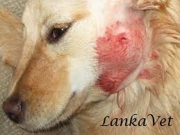
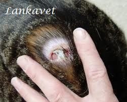
An abscess is a collection of pus encapsulated within a tissue underneath the skin. It occurs when the body tries to fight an infectious process usually caused by bacteria or parasites within the body tissues, sending white blood cells to fight further infection.
An abscess may first appear as a painful firm swelling under the skin that is hot or cold to the touch. In later stages, the localized swelling often becomes softer and less painful. This usually happens right before the abscess is getting ready to burst and drain.
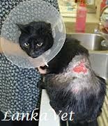
Abscesses around the neck and tail are far most common in cats who have access to the outdoors and who play territorial games with their fellow cats. Their teeth and claws are extremely contaminated with bacteria, and when they are sunk into the skin of the opposing cat, the bacteria becomes embedded under the skin. Pasteurella multocida is the most common bacteria to cause skin infections.

Causes

The most common causes of abscesses include:
  • Animal bites
  • Scratches
  • Trauma to the skin or underlying tissues
  • Foreign object penetration
  • Bad oral hygiene


How does it occur?

For an abscess to occur, the pet must have been injured somehow. They are caused by obstruction of oil (sebaceous) glands or sweat glands, inflammation of hair follicles, or minor abrasions and punctures of the skin. Germs get under the skin or into these glands, which causes an inflammatory response as your pet's natural defense mechanisms kick-in to remove the foreign body.
The middle of the abscess liquefies and contains dead cells, bacteria, and other debris. This area begins to grow, creating tension under the skin and further inflammation of the surrounding tissues. Pressure and inflammation cause the pain.
Pus is formed when the white blood cells die and accumulate in the affected area. As a protective mechanism, the immune system also works to wall off this area from the rest of the body, by forming a fibrous capsule. This fibrous capsule keeps the pus confined to one area. If the bacteria or foreign material overwhelms the immune system, the pus continues to accumulate and the abscess becomes larger.
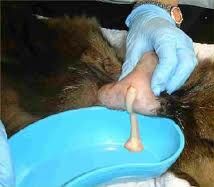
As time goes on, the wall surrounding the ever-growing pus pocket becomes thin, and at some point, it ruptures and pus drains out. Typically, the abscess ruptures to the exterior of the body, and pus drains from the wound. The pus is usually quite thick and creamy, and often has a distinct smell. However, in many cases; the abscess may not drain naturally and must be excised by a vet.

Signs
  • Swelling and inflammation
  • Heat and redness around the area
  • Pain around the area of infection
  • High temperature or fever
  • Lack of appetite
  • Lethargy and a reluctance to move
  • Rotting teeth, tooth loss, excessive saliva and bad breath (in mouth abscesses)
  • Hair loss (in skin abscesses)
  • Stiffness or lameness (abscesses around joints)
  • Licking or grooming affected area excessively
  • Foul odor
  • Purulent discharge (pus) from a wound

Treatment
  • Abscess drainage is painful and better to do it under anesthesia
  • Painkillers
  • Antibiotics to avoid secondary bacterial infections - Topical & intramuscular
  • Clindamycin - If it is serious or deep in the skin

Caring and Management
  • Clean hands thoroughly before handling abscess.
  • Clear the surrounding skin by clipping away hair with a small pair of scissors.
  • Gently clean the area with a mild soap and then rub it with clean gauze soaked with antibacterial solution, Ex. saline solution or hydrogen peroxide.
  • Gently pat the area dry with a towel.
  • Drain the abscess via surgical puncture and then drain with a sterile solution such as normal saline.
  • Abscesses are usually left open and allowed to drain to prevent the re-infection. 
  • Use a topical skin protecting agents, such as Betadine, to guard the skin against bacterial infection and keep the affected area clean.
  • Check it every 12 hours. Gently remove any scabs or crusts skin that have formed and clean the area with soap and water apply Betadine ointment.
  • If you come in contact with draining pus, just wash your hands. These bacteria may infectious to humans if it is exposed to an open wound on skin.